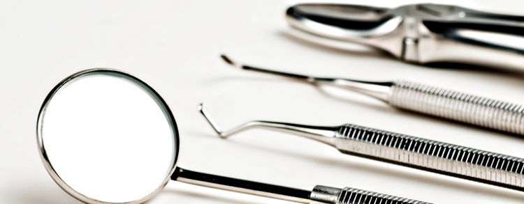
We use the latest and advanced equipment in all dental treatments. All the surgical instruments are sterilized and cleaned after every treatment.
A list of the equipment, technology and materials used in our clinic is given below.
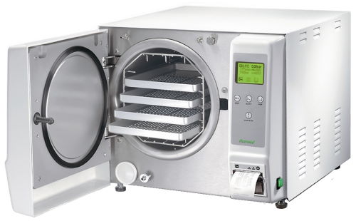
Sterilization Equipment
We use an automatic sterilization system of a Class-B Autoclave with an ultraviolet chamber.
Class-B Autoclave (Hot Air and Pressure Oven)
The surgical instruments are first sterilized in an autoclave machine at extremely high temperature and pressure. It kills all germs, bacteria, and viruses.
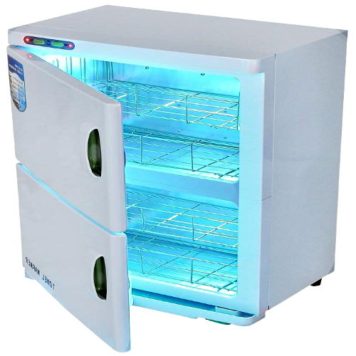
Ultraviolet Automatic Sterilization
After sterilization, the surgical instruments are then kept in an ultraviolet chamber, and are only taken out during a
treatment. An ultraviolet chamber keeps the sterilized surgical instruments under constant ultraviolet light. It prevents any new bacteria or virus to develop on the sterilized surgical instruments.
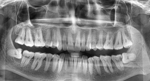
Digital X-Ray Devices
X-ray of Whole Mouth (OPG)
An x-ray of whole mouth shows the whole structure of the mouth. It shows all the teeth, blood nerves, veins, and condition of the jaw bone in the mouth. This x-ray is taken when the small x-ray of a single tooth is not sufficient, or when a major surgery is required.
Small X-ray of One Tooth (RVG)
The smaller x-ray device (RVG) takes a high resolution digital x-ray of a single tooth. After taking the x-ray, the image is visible on the computer screen.
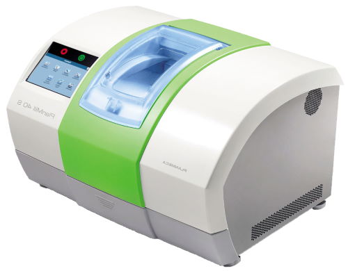
Equipment Used to Make Tooth Crowns, Dental Veneers, Dental Bridge
CAD / CAM Machine
A CAD / CAM (Computer Aided Design and Computer Aided Manufacturing) machine is used to manufacture tooth crown, dental veneers, Lumineers, and dental bridge from zirconia (ceramic) material. This machine has high accuracy and is completely automatic.
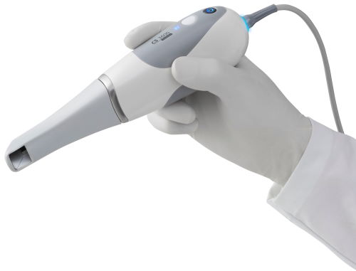
Intra-oral Scanner
An intra-oral scanner takes a 3D image of the mouth. This 3D image is directly sent to the CAD / CAM machine that makes tooth crown, dental veneers, Lumineers, and dental bridge from zirconia (ceramic) material, that fits perfectly on the tooth.
Equipment used in Dental Implant Surgery
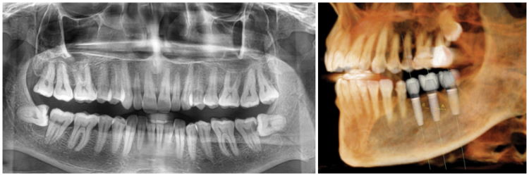
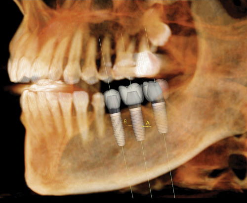
3D Image of Whole Mouth using CT Scan (CBCT) Machine
A CT scan (CBCT) machine gives a 3D image of your mouth. The 3D image gives the best locations in the jaw to insert a dental implant. Therefore, a very small hole is drilled in the gums to insert a dental implant because the exact location is already known to the dentist before cutting the gums.
If this 3D image of the mouth is not available, then a larger cut is required in the gums to find the best locations in the jaw to insert the dental implant.
The 3D image also gives information if any additional procedure is required, like: bone graft and sinus lift surgery. It also shows the path of main blood vessels in the mouth that must not be touched during the surgery.
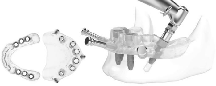
Stent Device to Insert Dental Implant
The dentist prepares a device called “Stent” using the 3D image of the mouth. The stent helps in accurate placement of the dental implant in the jaw bone. Therefore, a very small cut is required in the gums to insert the dental implant in the jaw.
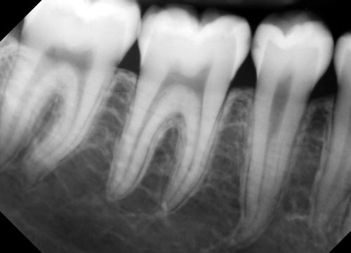
Equipment used in Root Canal Treatment
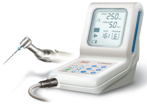
Endodontic Motor and Apex Locator
Endodontic motor and apex locator are automatic equipment that help in the complete removal of the infected tissue from the tooth.
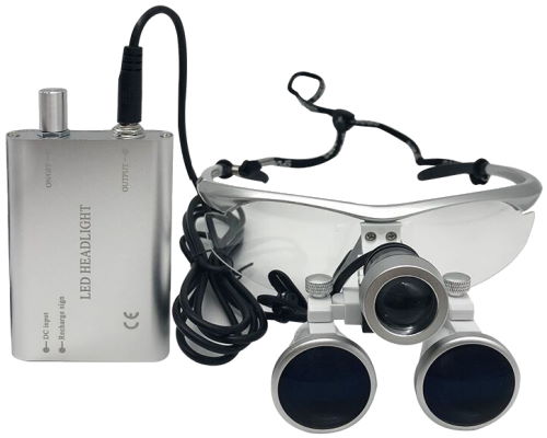
Dental Binoculars / Microscope / Loupes
A dental binocular (dental loupes) magnifies the tooth. It also has a bright flashlight attached to it. It gives better visibility of the tooth during the surgery.